Science
Can a Brain Have a Biological Age Different From Its Actual Age?
You are 45 years old. Your passport says so. Your birth certificate confirms it. But what does your brain say?
This is not a philosophical question. It is an increasingly precise scientific one, and the answer has implications for how we think about cognitive health, dementia risk, mental performance, and the ageing process itself.
The brain, as it turns out, does not age at the same rate as the calendar. Some people at 60 have brains that function with the biological vitality of a 45-year-old. Others at 45 show neural patterns more typical of someone two decades older. The gap between chronological age, the number of years you have been alive, and biological brain age, how old your brain appears to be at the cellular and structural level, is real, measurable, and meaningful.
Understanding why that gap exists, and whether it can be influenced, is one of the most active frontiers in neuroscience.
What Brain Age Actually Measures
Your chronological age is fixed, it moves forward at exactly one year per year, for everyone, without exception. Your biological brain age is different. It reflects the actual state of your neural tissue, the volume of different brain regions, the integrity of the white matter tracts that connect them, the efficiency of the metabolic processes keeping your neurons alive and communicating, and the molecular markers of cellular ageing within the brain itself.
Researchers measure biological brain age primarily through two lenses.
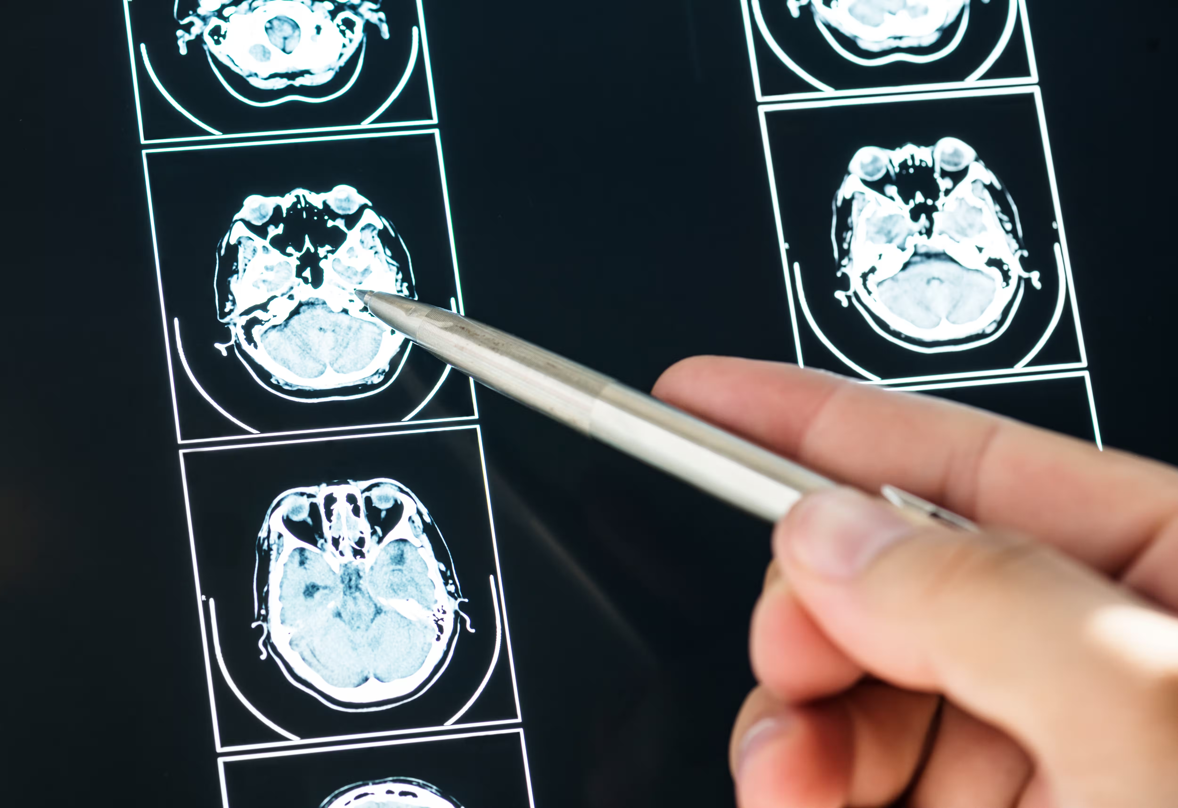
The first is structural MRI, brain scans that reveal the physical architecture of the brain with extraordinary precision. The brain changes in characteristic ways as it ages, grey matter volume decreases, particularly in the prefrontal cortex and hippocampus. White matter integrity declines. Ventricles enlarge as surrounding tissue is lost. These changes follow predictable trajectories that allow researchers to compare an individual's scan against population norms for their age, producing a brain age estimate that may be younger or older than their chronological age (Cole and Franke, 2017).
The second is epigenetic clocks, molecular tools that estimate biological age by measuring patterns of chemical modification to DNA. The most brain-specific of these clocks, developed from samples of neural tissue, estimate brain biological age independently of the rest of the body, and have found that the brain's biological age can diverge significantly from the biological age of other organ systems (Horvath, 2013).
Together, these tools have revealed something that should change how we think about brain health, the biological age of the brain is not fixed by the number of years you have lived. It is a dynamic property, influenced by genetics, but also profoundly shaped by the choices, exposures, and experiences of a lifetime.
The Brain-Age Gap and What It Predicts
A younger biological brain age, a brain that appears structurally and molecularly younger than chronological age would predict, is associated with a constellation of benefits that compound over time.
People whose brains age more slowly show better cognitive performance on tests of memory, processing speed, executive function, and reasoning, independent of their chronological age (Kaufmann et al., 2019). They show lower rates of dementia, including Alzheimer's disease, and when cognitive decline does eventually occur, it tends to begin later and progress more slowly. They show better mental health outcomes. And they show greater cognitive resilience following brain injury or illness, the younger-brained brain appears to have more reserve to draw on when things go wrong.
The opposite is equally true, and more alarming. People whose brains appear older than their chronological age are at elevated risk across all these dimensions. A study of more than 3,000 participants found that each year of brain age acceleration, each year by which biological brain age exceeded chronological age, was associated with meaningfully elevated risk of cognitive impairment, and that the gap was detectable years to decades before any clinical symptoms appeared (Franke and Gaser, 2019).
This is the clinical promise of brain age measurement, the possibility of identifying people at elevated risk of cognitive decline early enough for intervention to be meaningful before irreversible damage has accumulated.
What Accelerates Brain Ageing
Understanding what makes a brain age faster than it should is as important as understanding the measurement. The factors are multiple, and they interact.
Chronic stress is one of the most consistently documented accelerators of brain ageing. Sustained elevation of cortisol, the primary stress hormone, has toxic effects on hippocampal neurons, reducing their volume and impairing the synaptic plasticity that underlies memory and learning. Studies have found that people with high levels of chronic stress show greater grey matter loss in stress-sensitive brain regions, and that their brain age estimates are correspondingly elevated (McEwen, 2017).
Poor sleep is a second major accelerant. The brain's waste clearance system, the glymphatic system, which flushes out metabolic byproducts including amyloid-beta and tau proteins associated with Alzheimer's pathology, operates primarily during deep sleep. Chronic sleep deprivation impairs this clearance, allows toxic proteins to accumulate, and is associated with accelerated brain ageing in imaging studies (Xie et al., 2013).
Metabolic dysfunction, insulin resistance, obesity, type 2 diabetes, and chronically elevated blood glucose, drives neuroinflammation and impairs the energy metabolism of neurons, which are extraordinarily energy-hungry cells that depend on continuous glucose supply and mitochondrial function. People with type 2 diabetes show significantly accelerated brain ageing compared to metabolically healthy individuals (Moran et al., 2015).
Physical inactivity allows the brain to age faster. Exercise is one of the most robustly protective factors for brain health, it stimulates the production of BDNF (brain-derived neurotrophic factor), a protein essential for neuronal growth and survival, and it increases cerebral blood flow, reduces neuroinflammation, and appears to slow the structural changes associated with brain ageing across multiple imaging studies.
Loneliness and social isolation are associated with accelerated cognitive decline and brain ageing, an effect that is at least partly mediated through chronic stress and its neurological consequences. The social brain is a brain under continuous positive stimulation. Remove that stimulation, and the consequences are measurable.
Alcohol, smoking, and certain medications all have documented effects on brain structure and apparent brain age, the dose-response relationships well-established in imaging research.
What Slows, or Even Reverses, Brain Ageing
The encouraging dimension of this research is that the brain retains a degree of plasticity throughout life, the capacity to change, adapt, and recover, that means biological brain age is not simply a one-way ratchet. Some interventions appear to genuinely slow the rate of brain ageing. A small number appear to produce measurable rejuvenation.
Aerobic exercise is the intervention with the strongest and most consistent evidence. A meta-analysis of randomized controlled trials found that regular aerobic exercise increased hippocampal volume, one of the primary markers of brain ageing, in older adults, with effect sizes that correspond to reversing approximately one to two years of age-related hippocampal loss (Erickson et al., 2011). This is not a trivial finding. The hippocampus is the brain region most critically involved in memory and most vulnerable to age-related atrophy. Growing it, rather than losing it, is a meaningful intervention.
Sleep quality is perhaps the most modifiable and most undervalued brain protection factor. Prioritizing seven to nine hours of quality sleep per night, with consistent timing, a cool dark environment, and limited alcohol before bed, supports the nightly glymphatic clearance that removes the proteins implicated in neurodegenerative disease.
Cognitive engagement, intellectually demanding work, education, learning new skills, and maintaining complex mental activity, is associated with greater cognitive reserve and slower brain ageing. The "use it or lose it" principle has genuine neurobiological support; cognitive stimulation maintains synaptic density and appears to delay the expression of age-related structural changes.
Social connection is neurologically protective. The cognitive demands of social interaction, reading emotional cues, understanding context, maintaining relationships, provide continuous stimulation of the neural circuits that are most vulnerable to age-related decline.
Diet quality, particularly Mediterranean-style dietary patterns rich in polyphenols, omega-3 fatty acids, and diverse plant foods, is associated with slower brain ageing in large prospective studies, an effect mediated partly through cardiovascular health and partly through direct anti-inflammatory effects on neural tissue.
The Measurement Revolution, What Is Now Possible
The practical significance of brain age research has increased dramatically as the measurement tools have become more accessible and more precise.
Brain age estimation from MRI scans, using machine learning models trained on thousands of brain scans with known ages, has become a routine component of neuroimaging research, and is beginning to enter clinical practice. Tools that estimate brain age from a standard clinical scan are available in research settings and, increasingly, as commercial clinical tools.
More accessible still are blood-based biomarkers. Proteins shed by ageing or damaged neurons, including neurofilament light chain (NfL) and phosphorylated tau, can be detected in blood and provide indirect measures of brain health that correlate with imaging-based brain age estimates. As the sensitivity and cost of these blood tests improve, they are likely to become routine screening tools.
Epigenetic clocks derived from blood samples, while not as brain specific as those derived from neural tissue, provide estimates of biological ageing that correlate meaningfully with brain health outcomes. Several commercial longevity testing services now offer epigenetic age estimation alongside other biomarkers.
The convergence of these measurement tools, brain imaging, blood biomarkers, and epigenetic clocks, is creating the infrastructure for something that does not yet fully exist but is becoming increasingly plausible, routine, accessible brain health monitoring that tracks biological brain age over time and provides actionable feedback on whether it is accelerating or decelerating.
The Bottom Line
Your brain is ageing. That much is certain. But the rate at which it ages is not fixed, it is a dynamic property influenced by the choices, exposures, relationships, and habits of a lifetime.
Science is clear enough to be actionable. Exercise, consistently. Sleep, properly. Manage stress, actively. Stay socially connected. Eat a diet that supports brain health. Challenge your mind. Avoid the things that demonstrably accelerate neural ageing, chronic sleep deprivation, social isolation, metabolic dysfunction, heavy alcohol.
None of these is novel health advice. What is novel is the neuroimaging and molecular evidence that these interventions do not just make you feel better, they measurably slow the biological ageing of the most complex and irreplaceable organ you possess.
Your passport cannot be changed. Your brain age, the evidence increasingly suggests, can.
Cover image by Freepik [www.freepik.com]
References
Cole, J.H. and Franke, K. (2017) 'Predicting age using neuroimaging, innovative brain ageing biomarkers', Trends in Neurosciences, 40(12), pp. 681–690. doi,10.1016/j.tins.2017.10.001.
Erickson, K.I., Voss, M.W., Prakash, R.S., Basak, C., Szabo, A., Chaddock, L., Kim, J.S., Heo, S., Alves, H., White, S.M., Wojcicki, T.R., Mailey, E., Vieira, V.J., Martin, S.A., Pence, B.D., Woods, J.A., McAuley, E. and Kramer, A.F. (2011) 'Exercise training increases size of hippocampus and improves memory', Proceedings of the National Academy of Sciences, 108(7), pp. 3017–3022. doi,10.1073/pnas.1015950108.
Franke, K. and Gaser, C. (2019) 'Ten years of BrainAGE as a neuroimaging biomarker of brain aging, what insights have we gained?', Frontiers in Neurology, 10, article 789. doi,10.3389/fneur.2019.00789.
Horvath, S. (2013) 'DNA methylation age of human tissues and cell types', Genome Biology, 14(10), article R115. doi,10.1186/gb-2013-14-10-r115.
Kaufmann, T., van der Meer, D., Doan, N.T., Schwarz, E., Lund, M.J., Agartz, I., Alnæs, D., Barch, D.M., Baur-Streubel, R., Bertolino, A., Bettella, F., Boen, E., Borgwardt, S., Brandt, C.L., Buitelaar, J., Bürger, C., Cannon, D.M., Cattrell, A., Cooke, E.J., Coynel, D., D'Ambrosio, E., Dannlowski, U., Demro, C., Dhollander, T., Dima, D., Doan, N.T., Einevoll, G., Espeseth, T., Fatouros-Bergman, H. and Westlye, L.T. (2019) 'Common brain disorders are associated with heritable patterns of apparent aging of the brain', Nature Neuroscience, 22(10), pp. 1617–1623. doi,10.1038/s41593-019-0471-7.
McEwen, B.S. (2017) 'Neurobiological and systemic effects of chronic stress', Chronic Stress, 1. doi,10.1177/2470547017692328.
Moran, C., Phan, T.G., Chen, J., Blizzard, L., Beare, R., Venn, A., Munch, G., Wood, A.G., Forbes, J., Greenaway, T.M., Pearson, S. and Srikanth, V. (2015) 'Brain atrophy in type 2 diabetes, regional distribution and influence on cognition', Diabetes Care, 36(12), pp. 4036–4042. doi,10.2337/dc13-0143.
Xie, L., Kang, H., Xu, Q., Chen, M.J., Liao, Y., Thiyagarajan, M., O'Donnell, J., Christensen, D.J., Nicholson, C., Iliff, J.J., Takano, T., Deane, R. and Nedergaard, M. (2013) 'Sleep drives metabolite clearance from the adult brain', Science, 342(6156), pp. 373–377. doi,10.1126/science.1241224.
Test Your Knowledge!
Click the button below to generate an AI-powered quiz based on this article.
Did you enjoy this article?
Show your appreciation by giving it a like!
Conversation (0)
Cite This Article
Generating...