Science
Seeing Beneath the Skin - How Medical Imaging Helps Doctors Diagnose Without Cutting
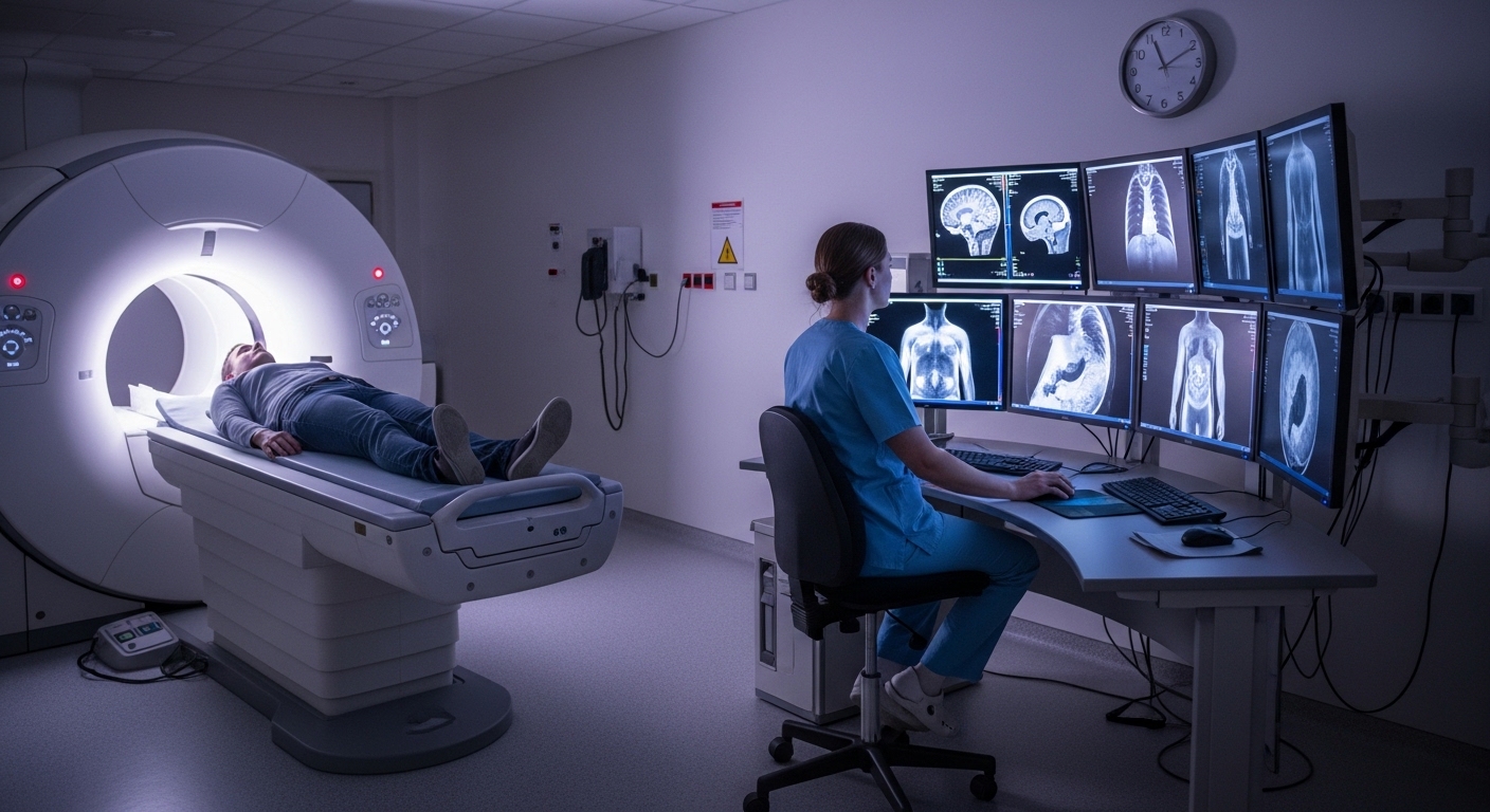
When a doctor orders an X‑ray, a CT scan, or an MRI, it can feel mysterious like your body is being turned into shadows and slices on a screen. But medical imaging is simply a set of tools that let doctors look inside without surgery. It shows bones, organs, blood vessels, and even how tissues are functioning, helping them find problems earlier, choose better treatments, and avoid unnecessary operations.
Modern diagnostics would be almost unthinkable without these technologies. A broken bone, a tiny stroke, a hidden tumor, a blocked artery imaging turns these invisible issues into visible evidence. Instead of guessing what might be wrong based only on symptoms, doctors can see the problem directly and act with much more confidence.
The Classics - X-Ray, CT, MRI, and Ultrasound
X‑rays are the oldest and simplest form of medical imaging. They use a small dose of ionizing radiation to create a 2D picture of dense structures like bones and teeth. That’s why they’re so common in emergency rooms and dental clinics, they are fast, cheap, and very good at showing fractures, infections in bone, and some lung problems.
CT scans (computed tomography) take X‑rays a step further. The scanner rotates around the body, capturing many images from different angles, then a computer reconstructs them into cross‑sectional slices. This allows doctors to see detailed views of organs, blood vessels, and complex fractures. CT is often used for stroke evaluation, trauma, and detecting tumors, but it uses more radiation than a standard X‑ray, so doctors weigh risks and benefits carefully.
MRI (magnetic resonance imaging) uses powerful magnets and radio waves instead of radiation. It is especially good at imaging soft tissues: brain, spinal cord, muscles, ligaments, and some tumors. MRI can show the difference between gray and white matter in the brain, detect multiple sclerosis lesions, and reveal subtle ligament tears that X‑rays would miss. The trade‑off is that MRI scans take longer, are noisier, and can feel claustrophobic for some patients.
Ultrasound uses high‑frequency sound waves to create images and does not involve radiation at all. A handheld probe sends sound into the body and listens for echoes, building a real‑time picture. It is widely used in pregnancy, to check the heart (echocardiography), and to look at organs like the liver, kidneys, and thyroid. Because it is portable and relatively inexpensive, ultrasound is a key tool in both large hospitals and smaller clinics.
Other modalities like PET and SPECT (nuclear medicine techniques) add another dimension by showing not just structure but function for example, how active a tumor is or how well the heart muscle is perfused. They do this by tracking small amounts of radioactive tracers inside the body.
From Pictures to Decisions - Diagnostics in Action
Imaging becomes diagnostics when those pictures are interpreted and used to make decisions. A chest CT might reveal a blood clot in the lungs, explaining sudden shortness of breath. A brain MRI can confirm whether slurred speech is due to a stroke, and if so, what type. An ultrasound of the abdomen might detect gallstones that match a patient’s pain pattern.
Doctors do not look at images in isolation. They combine them with the patient’s history, physical exam, lab tests, and sometimes genetic information. Imaging often changes the treatment plan: deciding whether surgery is needed, what kind of surgery to perform, or whether medication alone is enough. It is also vital for follow‑up monitoring whether a tumor is shrinking with treatment, or whether a fracture is healing correctly.
The AI Revolution - Computers That Help Read Scans
In recent years, artificial intelligence has joined radiologists in the reading room. Deep learning models can be trained on thousands or millions of labeled images to recognize patterns that are hard for the human eye to see. They can flag suspicious lung nodules on CT, detect early breast cancers on mammograms, or spot tiny brain bleeds on emergency scans.
AI tools are already being used to highlight abnormalities for radiologists, prioritize urgent cases in the worklist, and even generate draft reports. Studies suggest that AI can reduce diagnosis time by up to 30% in some workflows and improve consistency, especially for routine tasks. Importantly, these systems are designed to assist, not replace, human experts: the final decision remains with the radiologist or clinician, who considers the full clinical context.
Balancing Power and Risk
For all its benefits, medical imaging is not completely risk‑free. X‑rays and CT scans use ionizing radiation, which can slightly increase cancer risk over a lifetime, especially with repeated exposure. That’s why doctors follow the ALARA principle, As Low As Reasonably Achievable choosing the lowest radiation dose that still gives useful images and avoiding unnecessary tests.
Other modalities have different trade‑offs. MRI is generally safe but can be problematic for people with certain metal implants or severe claustrophobia. Ultrasound is considered very safe but is operator‑dependent the quality of images and their interpretation can vary with the skill of the person holding the probe. Nuclear medicine involves radioactive tracers, so timing, dosing, and disposal are carefully controlled.
Patients can and should ask questions: Why this scan? What will it show? Are there alternatives with less radiation or cost? In good practice, imaging is used when it will change management, not just to have a look.
Making Imaging More Accessible and Fair
One of the big challenges in global health is that advanced imaging is unevenly available. Large urban hospitals may have multiple MRI and CT scanners, while rural or low‑resource settings may rely mainly on basic X‑ray or portable ultrasound. New trends aim to close this gap: lower‑cost machines, mobile units, and AI‑assisted ultrasound that helps less‑experienced operators acquire and interpret scans more reliably.
At the same time, AI models must be trained and tested on diverse populations. If a model only learns from scans in one country or one demographic group, it may perform poorly elsewhere. Researchers are increasingly focused on building and validating systems that are fair and robust across different ages, ethnicities, and healthcare environments.
Medical imaging and diagnostics quietly sit at the heart of modern healthcare. They let doctors see beneath the skin, distinguish between look‑alike conditions, and track disease over time. With the rise of AI, these tools are becoming faster and more powerful, but also raise new questions about access, fairness, and the role of human judgement. For patients, understanding the basics what each scan does, why it’s ordered, and how it guides decisions can make the process less frightening and more empowering.
Test Your Knowledge!
Click the button below to generate an AI-powered quiz based on this article.
Did you enjoy this article?
Show your appreciation by giving it a like!
Conversation (0)
Cite This Article
Generating...